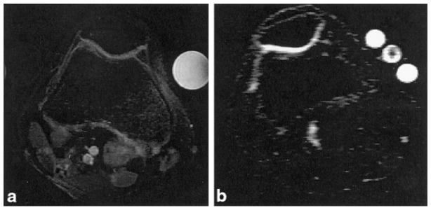
FIG. 4.
a: Axial proton fat-suppressed fast spin-echo image of a human knee joint. Two phantoms are included in the FOV: a water phantom (bright and right), and a fat phantom (dark and left). b: Axial 23Na MRI of the same knee joint. Three phantoms are included in the FOV. The center phantom is shaded because it is the end of the tube. The threshold was adjusted to yield maximum contrast between the cartilage and the surrounding tissue, thereby maximizing the signal intensities in the other two phantoms.

