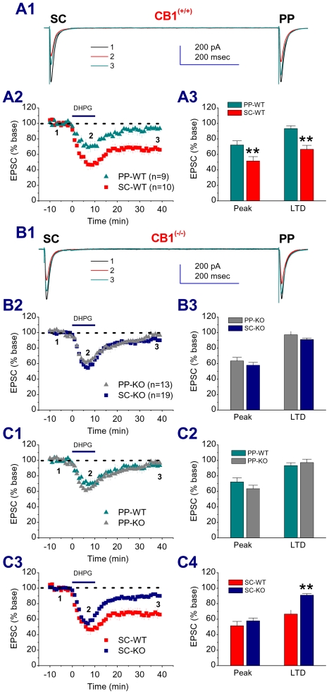
Figure 2. The difference in group 1 mGluR-induced LTD between the PP and SC pathways is absent in mice deficient in the CB1 receptor.
A1. Representative traces of EPSCs recorded in hippocampal CA1 pyramidal neuron from a wild-type (WT) mouse in response to independent PP and SC stimuli in the absence and presence of DHPG (50 µM) and washout. A2. Time courses of DHPG-induced changes in EPSCs. A3. Mean values of EPSCs averaged from 6 to 10 (Peak) and 36 to 40 min (LTD) following DHPG application. **P<0.01 compared with PP. B1. Representative traces of EPSCs recorded in a mouse from a CB1R knockout (KO) mouse in the absence and presence of DHPG (50 µM) and washout. B2. Time courses of DHPG-induced changes in EPSCs. B3. Mean values of EPSCs averaged from 6 to 10 (Peak) and 36 to 40 min (LTD) following DHPG application. C1. Time courses of DHPG-induced changes in EPSCs at the PP in CB1R KO and their WT littermates. C2. Mean values of PP EPSCs averaged from 6 to 10 (Peak) and 36 to 40 min (LTD) following DHPG application. C3. Time courses of DHPG-induced changes in EPSCs at the SC in CB1R KO and their WT littermates. C4. Mean values of SC EPSCs averaged from 6 to 10 (Peak) and 36 to 40 min (LTD) following DHPG application. **P<0.05 compared with WT.

