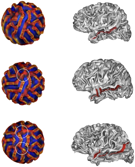
Figure 6. Three modes of variability and their correspondence on real anatomies.
First column: Three different modes of variability for the main fold observed on Fig. 5. We can see that the main sulcus, in one part at top left, is interrupted by a gyrus surrounded in white at middle left, and interrupted by two gyrus at bottom left. Second column: Two different modes of variability for the superior temporal sulcus on experimental data. Top: the superior temporal sulcus (STS) in pink is in one part. Middle: the STS is in two parts. Bottom: the STS is in three parts.

