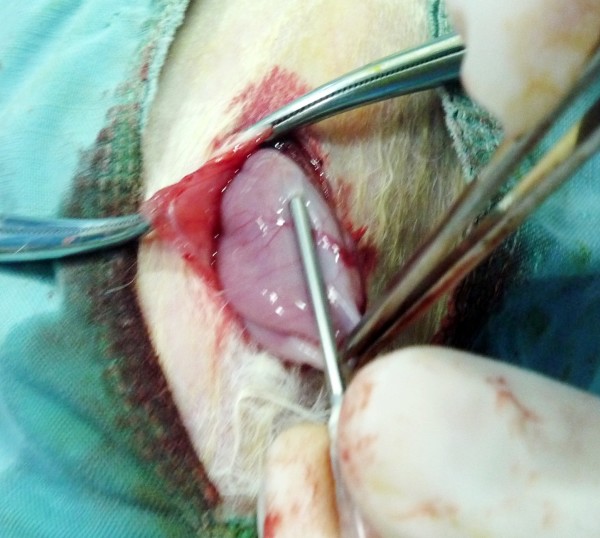
Figure 1.
Construction of rabbit peritoneal carcinomatosis model from gastric cancer. When the rabbit stomach was exposed under general anesthesia, a 16 G needle was inserted through the serosal and muscle layers into the submucosal layer of the stomach, and 0.1 mL of tumor cells (5×10¹⁰ vial cells/L) were injected, and injection site was pressed for 1 min to keep the tumor cells in place (See the text for detailed description).

