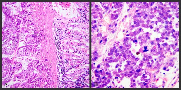
Figure 4.

Histopathology of rabbit model gastric cancer with PC, 2 weeks after VX2 carcinoma cells were injected into the submucosal layer of the stomach in Group A. VX2 tumor cells invaded the whole stomach wall (4A, ×100, HE stain) and showed many pathological mitotic figures (4B, ×200, HE stain).
