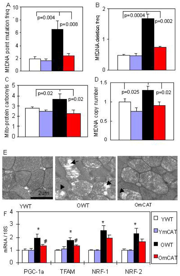
Figure 2. Mitochondrial DNA mutations, oxidative damage and biogenesis in aged heart and the protective effect of mCAT.
(A, B) Mitochondrial DNA point mutation and deletion frequencies (both per million base pairs) increased significantly with age and were attenuated in old mCAT mouse hearts. (C) Protein carbonyls content (nmol/mg) from cardiac mitochondrial extracts increased significantly with age, while mCAT attenuated age-dependent oxidative damage to mitochondrial proteins. (D) Mitochondrial DNA copy number (normalized to young WT) increased significantly with age, and old mCAT mice had significantly less increase in mitochondrial DNA copy number. (E) Electron micrographs displayed disruption of cristae and vacuolation of mitochondria (loss of electron density) in old WT mouse hearts, which was better protected in old mCAT heart mitochondria. (F) Upregulation of genes involved in mitochondrial biogenesis in the aged heart, including PGC-1a, TFAM, NRF-1 and NRF-2, and changes in PGC-1a and TFAM were attenuated in old mCAT mice (*P <0.025 between young vs. old WT, # P <0.025 between old WT vs. mCAT; n=9–12 each group; old: 26–28 months, young:4–5 months old).

