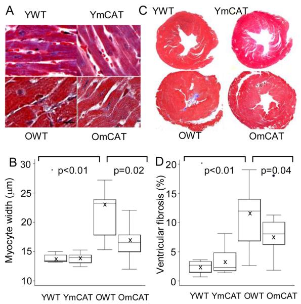
Figure 2. Cross-sectional study of cardiac pathology.
A) Old WT (OWT) mice had larger myocardial fiber width than old mCAT (OmCAT) mice (trichrome stain, 400x, scale bar:10μm). B) Quantitative analysis showed a significant increase in myocardial fiber-width in aged heart, which was significantly attenuated in OmCAT mice. C) OWT mice had more fibrosis (blue; trichrome , 20x) than OmCAT mice, as shown by quantitative image analysis of the percent fibrotic area in aged hearts, which was significantly attenuated in OmCAT mice (D).

