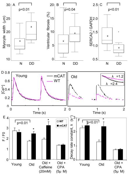
Figure 6. Plausible mechanisms of diastolic dysfunction in aged heart.
A) Myocardial fiber width was significantly greater in old mice with diastolic dysfunction (DD), compared with those with normal diastolic function (N). B) Ventricular fibrosis (%) was higher in old mice with diastolic dysfunction. C) SERCA2 protein was significantly decreased in mice with diastolic dysfunction. D) Representative calcium transients of isolated cardiomyocytes from young and old mouse hearts loaded with Fluo-4 and paced at 1 Hz. The inset illustrates OWT vs. OmCAT decay rates. E) Quantitative analysis showed significant decreases in Ca-transient amplitude (F/F0) and decay rate constant λ (s−1) in old WT, and protection in old mCAT cardiomyocytes. Sarcoplasmic reticulum Ca2+ load was also significantly higher in OmCAT cardiomyocytes, as shown by mobilization of Ca2+ by caffeine. The protective effect of mCAT was completely abolished by treating the old cardiomyocytes with cyclopyazonic acid, a SERCA-2 inhibitor.*p<0.025

