
Figure 6.
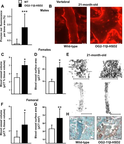
Preservation of bone lacunar-canalicular fluid, vascular volume, and vascular surface area in 21-month-old OG2-11β-HSD2 (black bars) compared to wild-type mice (white bars). Comparison of solute transport through the vertebral lacunar-canalicular system of male animals is shown in (A) and representative photomicrographs are shown in (B). Scale bars: 10 um. Comparisons of vertebral (C, D) and femoral (F, G) blood vessels after perfusion with lead chromate and representative μCT images from female animals (E). Scale bars: 1 mm. Data represent the mean ± SD. * P <0.05, ** P <0.02, *** P <0.002. (n = 6 to 11 per group. For measurements of procion red fluorescence and vertebral vessels, n = 3 per group). Representative photomicrographs of immunostaining for von Willebrand factor in sections of cancellous vertebral bone are shown in (H). The 21-month-old transgenics had an average of 77.8% greater vascular staining as compared with the similarly aged wild-type mice. Scale bars: 10 um. The arrow points to a longitudinal section of blood vessel.