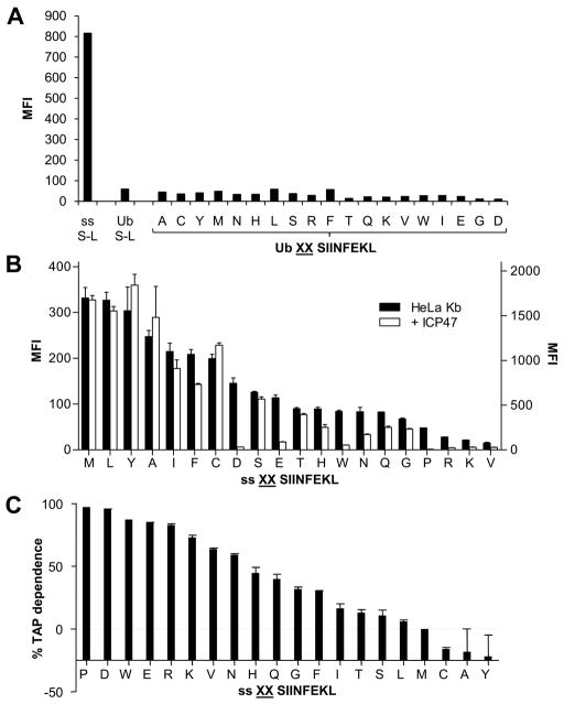
Figure 4. Affect of TAP inhibition on S-L presentation from cytosol and ER precursors.
HeLa-Kb-ICP47 cells were transfected with Ub XXS-L for 24hrs and stained for surface H-2Kb-S-L (A). ER targeted S-L (ss S-L) and cytoplasmic S-L (Ub S-L) were used as a positive control and negative control respectively. (B) Comparison of S-L presentation from ER targeted precursors following transfection of HeLa-Kb cells (black bars) and HeLa-Kb-ICP47 cells (white bars). Graph represents the average percentage presentation of two independent wells using the MFI of cells transfected with ss MMS-L as 100% presentation. Error bars represent the difference within each group. (C) Using the data in (B) the percentage TAP dependence of each ss XXS-L construct was calculated.

