
Figure 3b:
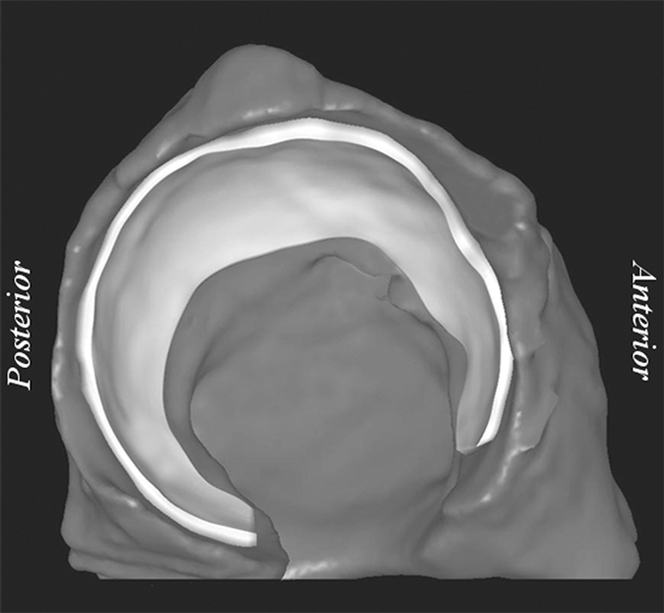
Representative right acetabulum. (a) Sagittal multidetector CT arthrogram shows 1.5-mm drill hole, cartilage, contrast agent, and subchondral bone (arrows). Note that drill hole was not filled with contrast agent, presumably owing to surface tension and high viscosity of the solution. (b) Lateral oblique 3D reconstruction of bone (dark gray) and cartilage (light gray) after semiautomatic segmentation.