Fig. 6.
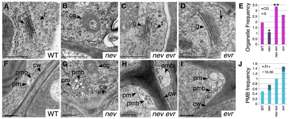
Mutations in EVR restore Golgi structure and location of the TGN in nev flowers. Transmission electron micrographs and analysis of cells in sepal AZ regions at the time of organ separation for wild-type (stage 16), nev (stage 16 non-shedding), nev evr (stage 15*) and evr (stage 16) flowers. (A-D) Instead of the flat stacks of Golgi cisternae characteristic of wild type (A), circularized multilamellar structures are observed in nev cells (B). Golgi with a wild-type appearance are found in nev evr (C) and evr cells (D). We frequently observed vesicular-tubular structures characteristic of the trans-Golgi network (TGN) (81%, n=16) closely associated with the Golgi cisternae (34±21 nm, n=13) in wild-type cells (A), whereas the TGN (15%, n=13) was less often observed near the circularized multilamellar structures (40±25 nm, n=2) of nev cells (B). The location of the TGN was restored in nev evr cells (83% associated with Golgi, n=24; 37±12 nm, n=20) and was unaffected by loss of EVR alone (76% associated with Golgi, n=21; 38±25 nm, n=15). (E) Frequency of flat Golgi cisternae (G, pink) and circularized multilamellar structures (CG, purple) per cell in sections of wild-type and mutant sepal AZ regions. For each genotype, n (cells)≥11. Statistical differences between nev and wild type, and between nev evr and nev tissues are indicated by single and double asterisks, respectively (Fisher's exact test, P<0.0001). A statistical difference was not detected between evr and wild-type tissues. (F-I) Paramural bodies (PMBs) were observed in the cells of wild-type (F), nev (G), nev evr (H) and evr (I) flowers. Whereas PMBs were observed in cells from each genotype, PMBs with greater than 30 vesicles were only observed in nev and evr cells. (J) Frequency of PMBs (10-30 and 31+ vesicles) per cell in sections of wild-type and mutant sepal AZ regions. For each genotype, n (cells)≥11. Statistical differences in PMB accumulation were not detected. cg, circularized multilamellar structures; cw, cell wall; g, Golgi cisternae; pm, plasma membrane; pmb, paramural body; t, trans-Golgi network. Scale bars: 0.5 μm.

