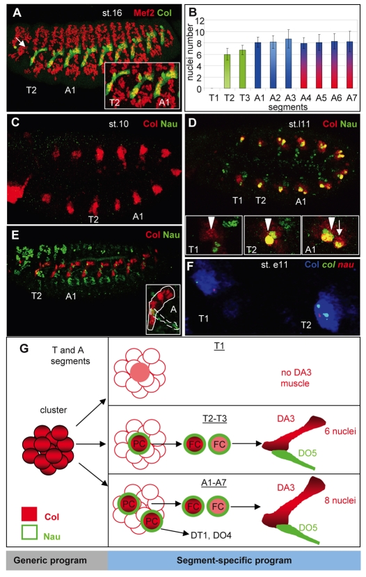
Fig. 1.
Segment-specific formation and properties of the DA3 muscle. (A) Mef2 (red) and Col (green) expression in a stage (st.) 16 Drosophila embryo, showing the entire muscle pattern and DA3 muscle, respectively. The arrow indicates the absence of DA3 muscle in the T1 segment. The inset shows an enlarged view of the T2, T3 and A1 segments. Note that the DA3 muscle is smaller in T than in A segments. (B) The segment-specific number of nuclei in the DA3 muscle (number of segments counted=30). The mesodermal domain of expression of Antp is in green, Ubx in blue and AbdA in red (Bate,1993) (see Fig. S3 in the supplementary material). (C-E) Comparison of expression of Col (red) and Nau (green) in stage 10 (C), late stage 11 (st.l11) (D) and stage 15 (E) embryos. Insets in D show the Col-expressing cluster in T1 (arrowhead), the DA3/DO5 progenitor in T2, and DA3/DO5 (arrowhead) and the DT1/DO4 progenitors (arrow) in A1. The inset in E is an enlarged view of the abdominal DA3 (circled) and DO5 (dotted circle) muscles that express Col and Nau, respectively. (F) Double in situ hybridization for col (green) and nau (red) primary transcripts and immunostaining for Col (blue). The T1 and T2 segments of an early stage 11 embryo are shown. Two dots per nucleus reflect the two alleles. All embryos are oriented anterior to the left and dorsal to the top. (G) Col (red) and Nau (green) expression during the process of DA3 muscle formation in different trunk segments, highlighting the segment-specific aspects. PC, progenitor; FC, founder cell.

