Fig. 1.
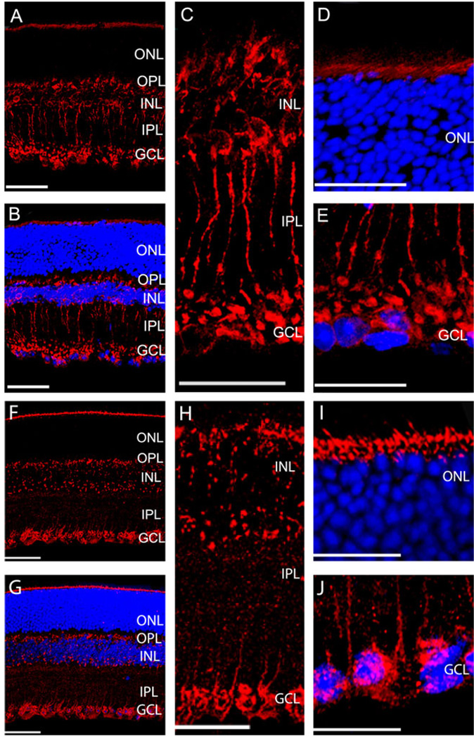
Expression of Rab1 (A–E) and Rab6 (F–J) in the transverse sections of the mouse retinas. (A and F) Representative immunostaining with Rab1 (A) and Rab6 (F) antibodies. (B and G) Immunostaining of Rab1 (B) and Rab6 (G) antibodies (red) and nuclear staining of DAPI (blue). (C and H) Enlarged sections of immunostains from the INL to the IPL with Rab1 (C) and Rab6 (H) antibodies. (D and I) Enlarged sections of immunostains from the outer border of ONL with Rab1 (D) and Rab6 (I) antibodies. (E and J) Enlarged sections of immunostains from the GCL with Rab1 (E) and Rab6 (J) antibodies. ONL, outer nuclear layer; OPL, outer plexiform layer; INL, inner nuclear layer; IPL, inner plexiform layer; GCL, ganglion cell layer. Scale bar is 50 µm in (A, B, F, and G), and it is 25 µm in (C–E) and (H–J).

