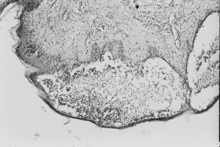
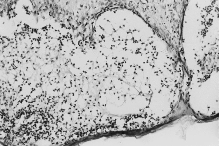
Figure 2.
A-B. Histopathology findings of pemphigus vulgaris.
Suprabasal intraepidermal blister, subtle spongiosis with no marked evidence of acantholysis, at the edge of the blister is focal collections of eosinophils and lymphocytes. In the papillary dermis scattered lymphocytes and eosinophils are present. This appearance is known as eosinophilic spongiosis.


