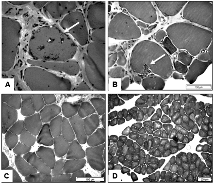
Figure 4.
Muscle biopsy findings in a 61 year old man with a 20 year history of untreated IBM. A and B: From a vastus lateralis biopsy showing chronic changes with interstitial fibrosis, myofibre atrophy and a fibre with rimmed vacuoles (arrow), and sparse inflammation (A); invasion of a non-necrotic muscle fibre by mononuclear cells (arrow) (B). C and D: From a deltoid biopsy showing a ragged-red fibre (C) and multiple blue-staining COX-negative fibres (D).

