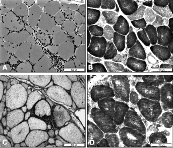
Figure 5.
Biopsies from deltoid (A and B) and biceps muscles (C and D) in a 35 year old male with a 3 year history of sIBM showing endomysial mononuclear inflammatory infiltrates and myofibre invasion (A); diffuse MHC-1 upregulation (C); and multiple blue-staining COX-negative fibres in both muscles, which are more atrophic in the biceps muscle (B and D). Fibres with rimmed vacuoles were present but were sparse in both muscles.

