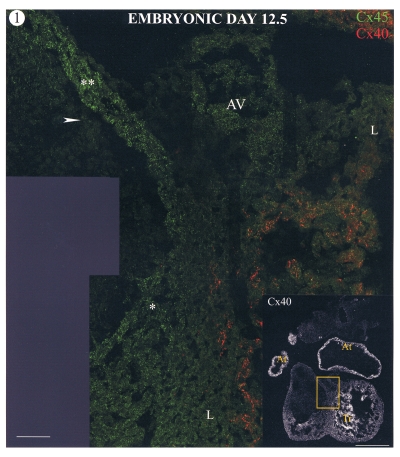
Figure 1.
Confocal images of a section through a mouse heart at embryonic day 12.5. The low power image (insert; bar marker = 500 μm) shows connexin40 (Cx40) labelling, which is present in the atrial tissue (At) and trabeculations (Tr). The boxed area is shown at high magnification double labelled for connexin45 (Cx45) (green) and Cx40 (red). Regions of high Cx45 labelling are restricted to the conus myocardium of the outflow tract (**) and atrioventricular junctional (AV) and interventricular septal myocardium (*), which have more intense labelling than other regions (L). Cx45 label is also present in the cushion mesenchyme adjacent to the conus myocardium (arrow). Bar marker = 50 μm

