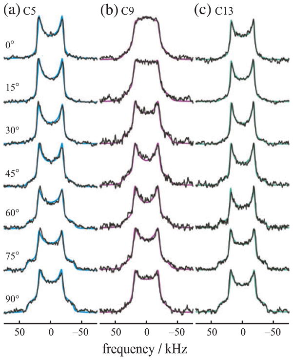
Figure 2.
Orientation-dependent 2H NMR spectra for aligned rhodopsin POPC (1:50) recombinant membranes provide angular restraints for retinylidene ligand in the dark state. (a–c) 2H NMR spectra for 11-Z-[5-C2H3]-retinylidene rhodopsin (blue), 11-Z-[9-C2H3]-retinylidene rhodopsin (magenta) and 11-Z-[13-C2H3]-retinylidene rhodopsin (green) at pH 7 and T = −150°C. Theoretical lineshapes for an immobile uniaxial distribution (solid lines) are superimposed on the experimental 2H NMR spectra. Note that characteristic lineshape changes are observed as a function of the tilt angle, which manifest the different methyl bond orientations with respect to the membrane frame. Reproduced with permission from Struts et al. (64).

