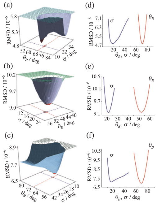
Figure 3.
Global fitting of 2H NMR spectra for 11-cis-retinal in the dark state of rhodopsin gives methyl bond orientations and mosaic spread of aligned membranes. (a–c) RMSD of calculated versus experimental 2H NMR spectra for retinal deuterated at C5-, C9- or C13-methyl groups, respectively and (d–f) cross-sections through hypersurfaces. Distinct minima are found in the bond orientation θB and mosaic spread σ of aligned membranes. Reproduced with permission from Struts et al. (64).

