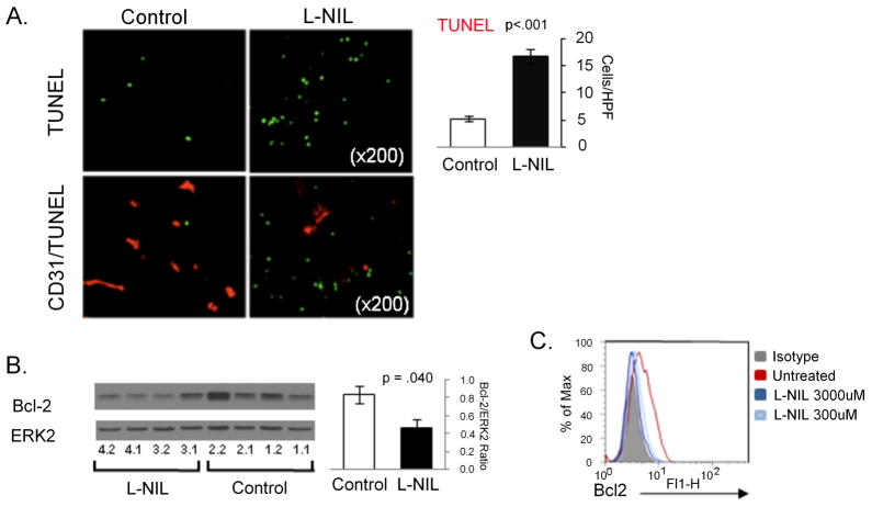
Figure 5. L-nil downregulates Bcl-2 expression and induces intratumoral apoptosis in vivo.
A. Mel624 xenografts were harvested from mice after treatment with 0.15% L-nil or plain water control for 14 days. Paraffin-embedded slides were subjected to TUNEL analysis of apoptotic cells (top), or double-staining for TUNEL and the vascular marker CD31(bottom). Representative images are shown on the left and quantitation of TUNEL staining on the right. B. Western blot analysis mel624 xenograft lysates was performed to confirm downregulation of Bcl-2 expression in tumors from L-nil treated mice (2 replicates of 2 tumors per group). The bar graph shows quantitation of Bcl-2 levels (normalized to Erk2 protein expression) in tumors from control and L-nil treated mice. C. 5×10⁴ A375 cells were cultured in 48 well plates for 3 days with the indicated concentration of L-nil, at which time the medium and inhibitor were replenished and cells cultured for an additional 2 days prior to immunostaining for intracellular Bcl-2 and analysis by flow cytometry.

