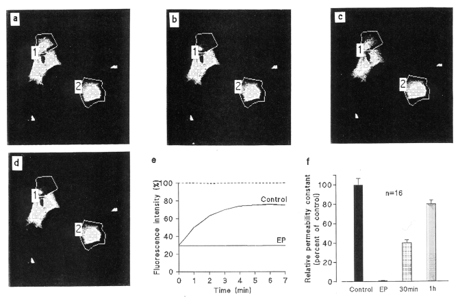
Figure 2.
Example of interruption of cell-to-cell dye diffusion elicited by 17β-estradiol propionate (EP) in ventricular myocytes of neonatal rats in primary culture. The grey density images of fluorescence intensities were obtained by scanning a 140×140 μm field with low intensity light pulses. After a prebleach scan (a), 6-carboxyfluorescein (6-CF) was photobleached in some selected areas (polygon 1) by means of strong illumination. The fluorescence levels in the selected cells were recorded just before (a) and just after photobleaching (b), and again 7 min later in control conditions (c) then in the same cells exposed to 17β-EP (25 μM) for 15 min (d). The graphs (e) show a comparison of the time courses of the fluorescent emission of the selected cells in control conditions (Control) and after treatment with EP. The fluorescence intensity is represented as a percentage of the prebleach emission versus the time after photobleaching. In control conditions, the fluorescence emission of the bleached cell increased progressively while, in contrast, it did not significantly change cells that were exposed to the steroid. The unbleached cell in polygon 2 served as a control. (f) The relative permeability constant k was reduced to immeasurably low values after a 15 min exposure to the steroid, then progressively increased after washing, when fluorescent dye diffusion became possible from neighbouring cells through reopened junctional channels

