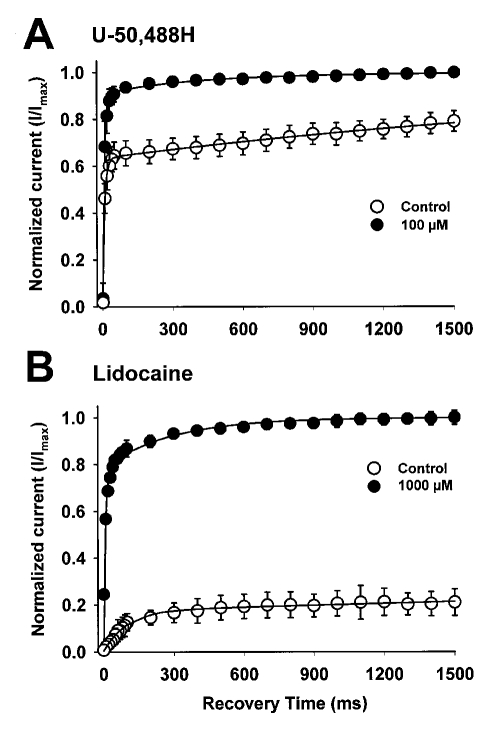
Figure 5.
Effects of 100 μM U-50,488H (A) and 1000 μM lidocaine (B) on recovery from inactivation of rat heart sodium channels. Oocytes were held at −120 mV and depolarized with an inactivating pulse to −10 mV for 500 ms. This was followed by a variable recovery interval ranging from 10 to 1500 ms at −120 mV. The recovery interval was followed by a 20 ms test depolarization to −10 mV. The peak current amplitude elicited during the test pulse was normalized to the peak current amplitude during a 22 ms pulse elicited from a holding potential of −120 mV for the rat heart channel immediately before the recovery protocol. The mean ± SD for the normalized current is shown as a function of the recovery time for at least four oocytes either in the absence of drug or in the presence of 100 μM U-50,488H (A) or 1000 μM lidocaine (B). The smooth curves represent fits of the data to a double exponential equation

