Figure 1.
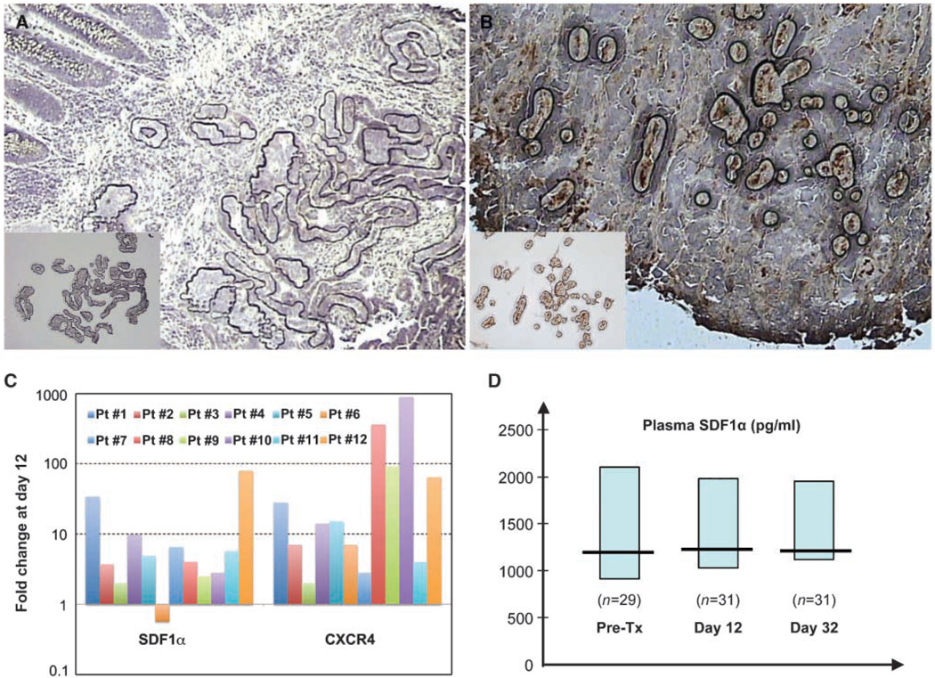
Changes in expression in cancer cells and TAMs from rectal carcinoma and in circulating plasma SDF1α levels after bevacizumab treatment. A, representative image of tumor tissue after selected tumor cells were burned by the capture laser and before RNA extraction (inset). B, selection of TAMs for LCM guided by CD68 immunostaining. Images of TAMs, burned by the capture laser of the cap containing TAMs (inset ; magnification, ×20). C, comparison of relative SDF1α and CXCR4 RNA levels in tumor cells captured by LCM from 12 pairs of serial biopsy samples collected before and 12 d after bevacizumab monotherapy. Columns, mean fold change in gene expression compared with pretreatment value. GAPDH was used as RNA control in the PCR. D, kinetics of plasma SDF1α after treatment with bevacizumab alone (day 12) and after bevacizumab with chemoradiation (day 32). Note the relatively high circulating levels of SDF1α (median of >1 ng/mL, shown with 95% confidence intervals) and the lack of consistent change in the overall population. Pre-Tx, pretreatment.

