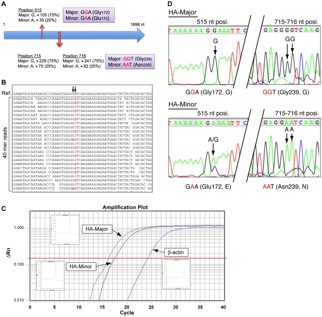
Figure 3. Genetic variations of the HA nucleotide sequence.
(A) Schematic representation of 3 nucleotide variations (positions 515, 715, and 716 nt) in the HA coding nucleotide sequence. Three variations were classified as Major (75% appearance) or Minor (25% appearance) by read coverage (×), and the coding amino acids are also shown. (B) Arrows indicate positions 715 and 716 nt of the HA sequence, and the alignment image of the 40-mer reads. Nucleotides shown in red are the mismatches to the reference sequence of A/Tronto/T0106/2009(H1N1). Every read suggested that either the GGT or AAT sequence was present, but not the GAT or AGT sequence. (C) An amplification plot for HA-specific qRT-PCR. (D) Validation of genetic variation by Sanger capillary sequencing. HA-Major or HA-Minor PCR products were obtained by qRT-PCR using HA-Major- or HA-Minor-specific PCR primers. HA-Major PCR product shows G nucleotides at positions 515, 715, and 716 nt, while HA-Minor shows A nucleotides.

