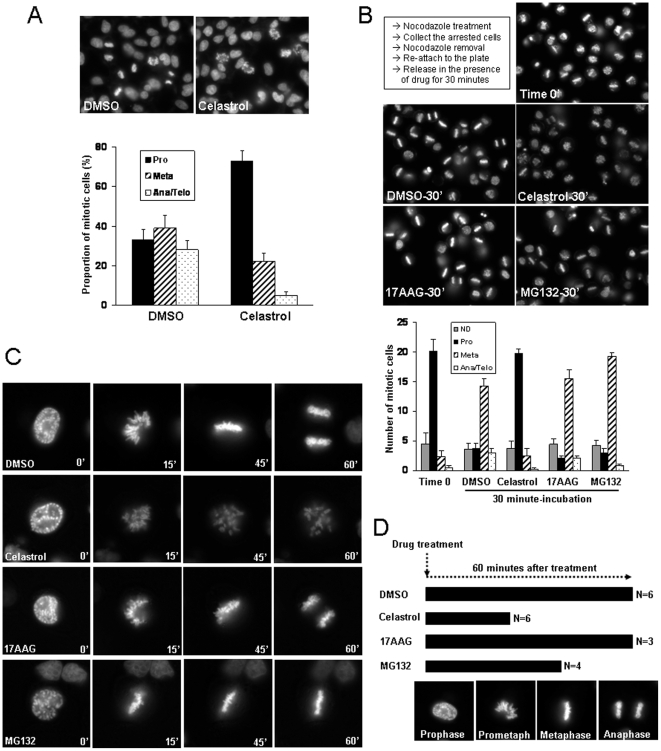
Figure 2. Celastrol inhibits mitotic progression and chromosome alignment.
(A) HEK293 cells were treated with Celastrol (2 µM) for 1 hour before fixation and staining with DAPI. The mitotic cells were counted and scored according to their chromosomal morphology. The proportion of each mitotic stage over the total number of counted cells was presented. (B) HeLa cells were treated with Nocodazole (100 nM) for 6 hours. The arrested cells were collected by tapping the plate, washed in PBS, and replated in a serum free medium. After 15 minutes of attachment (time 0), each drug was added and incubated for 30 minutes before fixation and staining with DAPI. The average number of cells in each stage from the quadruplicates was shown. ‘ND’ indicates the non-mitotic or dead cells. (C) Hela cells over-expressing EGFP-H2B protein were treated with indicated drugs. Immediately following drug addition, the prophase cells were identified and the time-lapse images were taken every 5 minutes for 90 minutes. The magnified still images from the representative movies were shown. (D) The summary of time-lapse movies for mitotic chromosome alignment in C). The degree of mitotic progression of prophase cells in 60 minutes in the presence of each chemical was determined based on chromosomal shape, and was indicated by horizontal bar. Numbers indicate number of cells examined. The representative chromosomal shapes of control cells at different mitotic stages were shown in lower panel as a reference.

