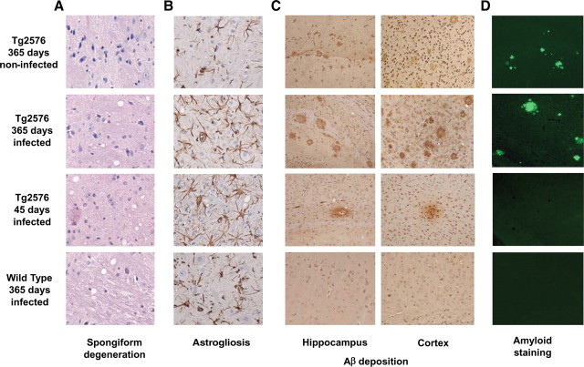
Figure 2.
A–D, Brain histopathological studies. Representative animals from different groups were studied histopathologically for spongiform brain degeneration after hematoxylin–eosin staining (A), reactive astrogliosis by GFAP staining (B), Aβ deposition by immunohistochemistry using the 4G8 anti-Aβ antibody (C), and staining with the amyloid-specific dye thioflavin S (D). It is important to emphasize that the prion deposits in mice affected by RML prions are not thioflavin S positive but are rather diffuse prefibrillar aggregates. The images in A and B correspond to the medulla, C to the hippocampus or cortex as indicated, and D to the cortex. (See also supplemental Figs. 2, 3, available at www.jneurosci.org as supplemental material.)

