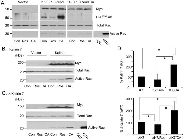
Fig. 7. The GEF activity of Kalirin is increased following Calyculin A treatment of cells.
Activation of Rac was assessed using GST-Pak-Crib. A. pEAK RAPID cells transfected with parent vector or vectors encoding KGEF1→7end or KEGF1→7end-Thr1590/Ala1590 were untreated (Con) or treated with 10μM roscovitine (Ros) for 4h or 25nM Calyculin A (CA) for 30min. Transfected proteins were detected with myc antibody (upper); phosphorylated protein was detected with P-T1590 Ab (second), and total Rac (third) was visualized with Rac antibody. GTP-bound Rac was isolated using GST-Pak-Crib resin and quantified by Western blot analysis with Rac antibody (bottom). B and C. pEAK RAPID cells transfected with vectors encoding Kalirin7 (B) or μKalirin7 (C) were analyzed as described in A. Control cells transfected with parent vector. Pooled cell extract incubated with GDP or GTPγS, negative and positive controls. D. Data from three independent assays quantified; activated Rac in control cells expressing Kalirin7 (upper) or μKalirin7 (lower) set to 100%; activated Rac in roscovitine or Calyculin A treated cells expressed as a percentage of relevant control; p values calculated using Student’s T test; *, p<0.05.

