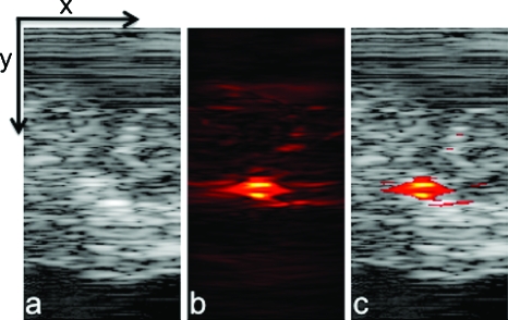
Figure 4.
(a) Ultrasound, (b) photoacoustic, and (c) combined images of nanoparticles injected directly into an ex vivo canine pancreas. All images were acquired from the same position as determined by the location of the ultrasound transducer. The images are 20 mm deep (y axis) and 10.5 mm wide (x axis).

