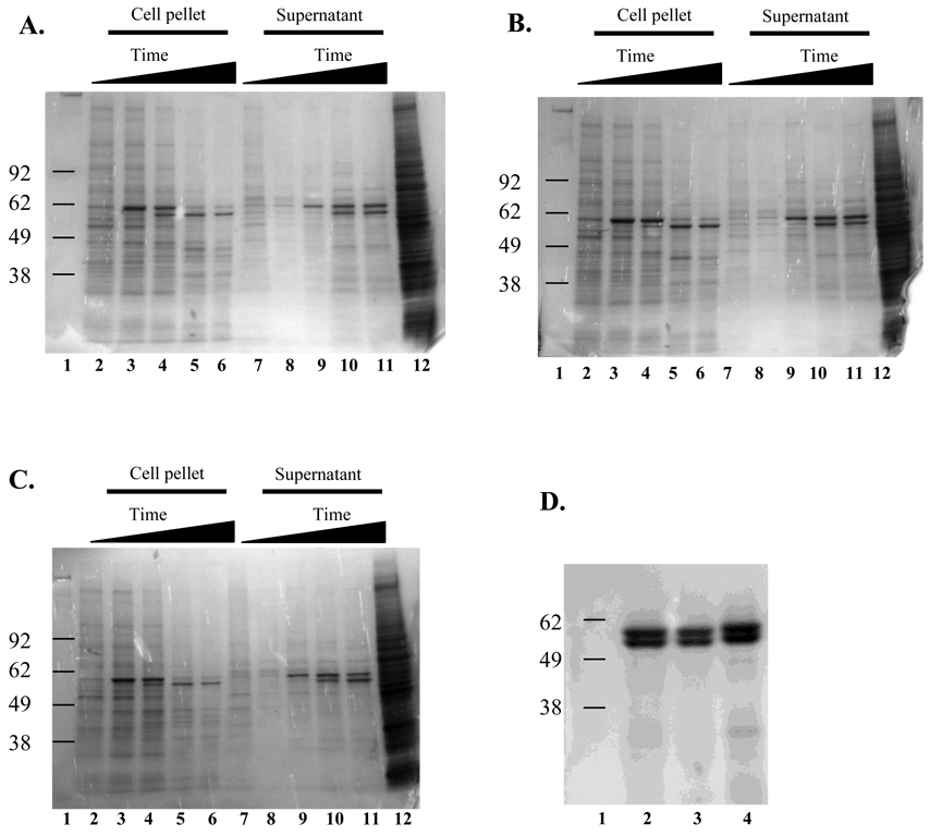
Fig. 1.
SDS-PAGE analysis of SSL vesivirus proteins produced in the baculovirus expression system. Proteins were separated by SDS-PAGE and stained with Gel Code Blue (Invitrogen). Panels A–C: samples were harvested daily for 5 d from recombinant baculovirus infected Sf-9 cell cultures. Lane 1: molecular weight marker, lanes 2 through 6: daily collections from cell pellets, lanes 7 through 11: daily collections from cell culture supernatant, lane 12: uninfected Sf-9 cell control. Panel A) V810 VP1, Panel B) V1415 VP1, Panel C) V810 VP1+VP2, Panel D) proteins purified from cesium chloride gradients of the supernatant from day 5 from the 3 recombinant viruses. Lane 1: molecular weight marker, lane 2: V810 VP1, lane 3: V1415 VP1, lane 4: V810 VP1+VP2.

