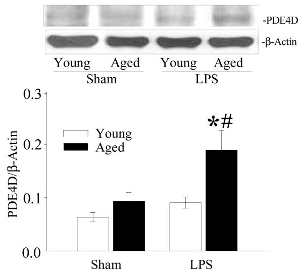
Figure 5. Increased expression of PDE4D in splenic tissues of septic aged rats.
Proteins were extracted from splenic tissues from sham and LPS exposed young and aged rats and examined for PDE4D protein by Western blotting. β-actin antibody was used to correct for changes in protein loading. Data are shown as PDE4D/β-actin ratio. Data are presented as mean ± SE (n=6) and compared by two-way ANOVA and Student-Newman-Keuls method: * p<0.05 versus respective sham; #p <0.05 versus young sham or LPS.

