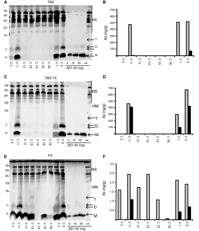
Figure 3.
Aβ monomer and SDS-stable dimer are detected in cortical samples serially extracted with Tris-buffered saline (TBS), Tris-buffered saline containing 1% TX-100 (TBS-TX) and formic acid. Aliquots of nine brain samples (0.2 g, temporal cortex) were serially extracted and analysed. Western blots of TBS (A), TBS-TX (C) and formic acid (E) extracts are shown. NS indicates non-specific immunoreactive bands detected in buffer alone (−ve) and molecular weight markers are indicated on the left. The concentration of Aβ present in each extract was determined by comparison with the synthetic Aβ standards shown and estimated using Li-COR software (B–F). M = monomer; D = dimer; T = trimer; HM = high molecular weight Aβ species larger than trimer; FA = formic acid.

