
Fig. 1.
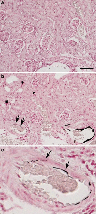
Light micrographs of sections of the kidney cortex of a wild-type (a) and an Abcc6 −/− mouse (b, with detail in c), stained for calcification with von Kossa. The mice were killed after 12 months of a diet supplemented with Ca (4×Ca diet). Extensive calcification (black deposits) is present in arteries in the kidney cortex of the Abcc6 −/− mouse (b). Please notice that the calcification is located within the wall of the artery (arrows in b and c, which shows a detail of b). Bar represents 100 μm