Fig. 4.

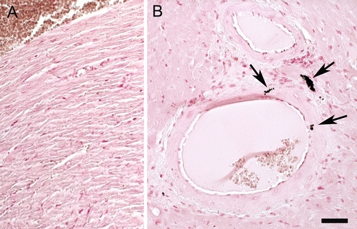
a Micrographs of paraffin sections of the ventricle of the heart of a wild-type (a) and an Abcc6 −/− mouse (b). Von Kossa staining demonstrates calcifications (black deposits; arrows) in walls of arteries in the Abcc6 −/− mouse. Bar is 50 μm

a Micrographs of paraffin sections of the ventricle of the heart of a wild-type (a) and an Abcc6 −/− mouse (b). Von Kossa staining demonstrates calcifications (black deposits; arrows) in walls of arteries in the Abcc6 −/− mouse. Bar is 50 μm