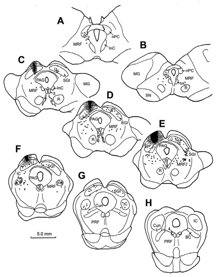
Figure 1.

Chartings of the distribution of retrogradely labeled neurons (dots) in the rostral brainstem following a BDA injection into the superior colliculus (C–F) in the cat. A rostral to caudal series of frontal sections is shown in this and other chartings. The core of the injection site, and most of the surrounding corona (stipple) were contained within the superior colliculus and involved all layers. Retrogradely labeled neurons are located in the contralateral intermediate gray layer (SGI) (C–G). Labeled neurons are also present ipsilaterally in the nucleus of the posterior commissure (nPC)(A&B), bilaterally in the dorsal portion of the midbrain reticular formation (MRF) (A–F) and bilaterally in the rostral pole of the inferior colliculus (IC)(F&G), and the cuneiform nucleus (G&H).
