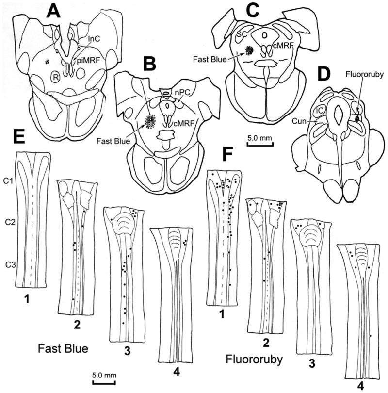
Figure 10.

Chartings demonstrating the distribution of retrogradely labeled neurons (dots) in the cervical spinal cord following injections of the cMRF (left) and cuneiform nucleus (right) of a macaque. The distributions of the cells are shown in two dorsal (D) to ventral (V) chartings of the same series of horizontal sections through the upper cervical spinal cord (E1–4 and F1–4), that were made for each fluorophore. Fast Blue, injected into the physiologically defined left cMRF (A–C) retrogradely labeled neurons primarily in the ipsilateral ventral horn of the spinal cord (E2&3). Fluororuby, injected into the right cuneiform nucleus (D), retrogradely labeled neurons primarily in the ipsilateral dorsal horn (F1).
