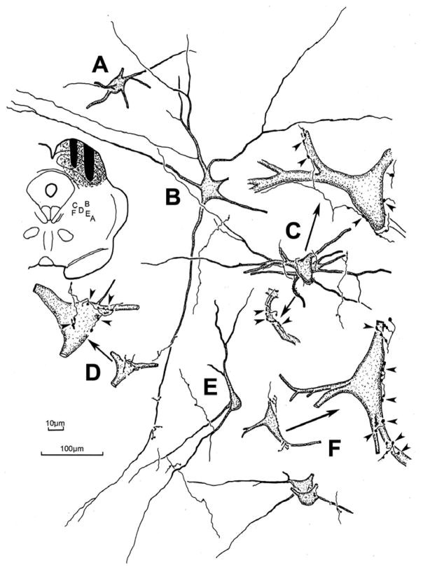
Figure 3.
The morphology of the labeled reticulotectal neurons and associated labeled tectoreticular axons in the cat. The BDA injection site and location of the illustrated examples is shown in the charting in the upper left. The labeled neurons were multipolar in shape and, in some cases (C,D&F), displayed numerous close associations between the labeled boutons of individual axons and a labeled cell. Other examples (A,B&E) showed few or no close associations. Arrows in C,D&F point to higher magnification drawings of examples with close associations (arrowheads). 100μm scale bar for cells A–F; 10 μm scale bar for enlargements of C,D&F.

