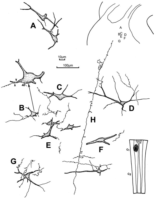
Figure 6.
The morphology of the labeled reticulospinal neurons and associated labeled spinoreticular axons in the cMRF of the cat. The BDA injection site is illustrated in the lower right and the location of the illustrated examples is shown in the upper right. The labeled neurons were multipolar in shape and, in some cases (B,E&G), displayed numerous close associations between the labeled boutons of an individual axon and a labeled cell. Other examples showed only a few (C–F) or no (A) close associations. Arrow points to a higher magnification drawing of an example with close associations (arrowheads) in B. The spinoreticular main axons (H) often had long dorsoventral courses, with mediolaterally running branches. Axon H was associated with neurons D&F. 100μm scale bar for cells A–G; 10 μm scale bar for enlargement of B.

