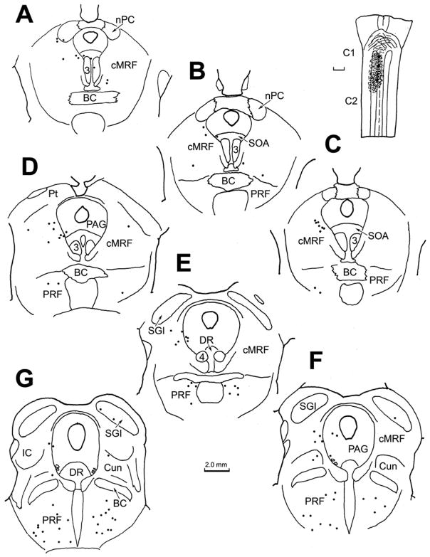
Figure 8.
Chartings demonstrating the distribution of retrogradely labeled neurons following an injection of BDA into the spinal cord of a macaque at the level of C1. The injection site is illustrated in a longitudinal section in the upper right. Retrogradely labeled neurons (dots) were found in both the midbrain and pontine reticular formation. The labeled cells were primarily ipsilaterally distributed in the cMRF (A–F) and bilaterally distributed in the pontine reticular formation (C–G). The cMRF cells were mainly located medially, and some labeled cells were found in the adjacent periaqueductal gray. The scale bars indicate equivalent distances.

