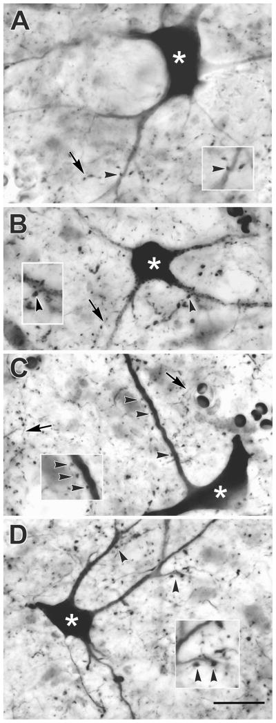
Figure 5.
Photomicrographs of anterogradely labeled reticulotectal axon arbors (arrows) and retrogradely labeled tectoreticular neurons (asterisks) in monkey A. Examples from the upper (A&B) and lower (C&D) sublaminae of SGI are shown. Close associations (arrowheads) were present between the boutons of labeled axons and the dendrites of tectoreticular neurons are shown at higher magnification in inserts. Scale = 25 μm, 40 μm for inserts.

