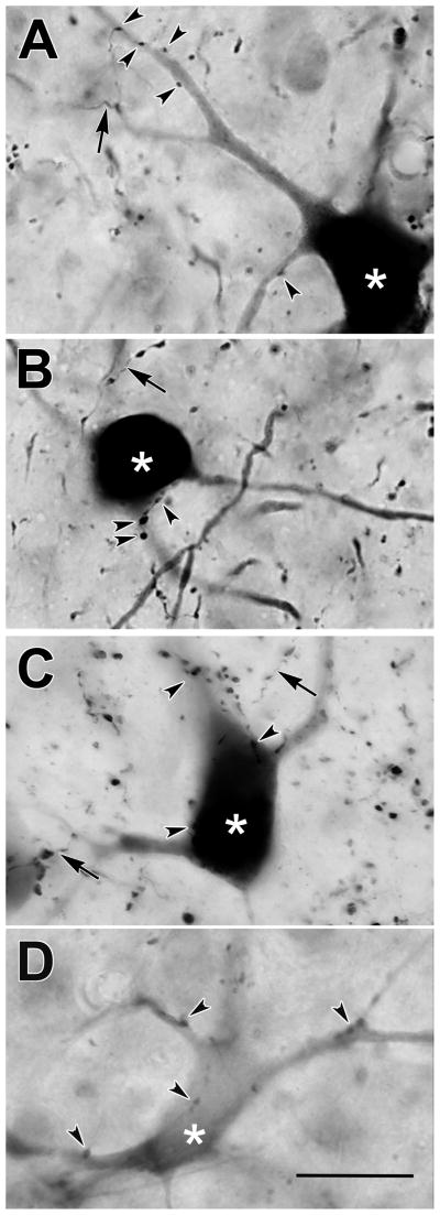
Figure 7.
Photomicrographs of retrogradely labeled tectoreticular neurons (asterisks) and anterogradely labeled reticulotectal axonal arbors (arrows) in monkey B. The labeled neurons located in the upper sublamina of SGI (A–C) were often well labeled with BDA, while examples from the lower sublamina of the SGI (D) were generally much more lightly labeled. Close associations (arrowheads) were present between some reticulotectal boutons and tectoreticular neurons in both sublamina (A–D). Scale = 25 μm.

