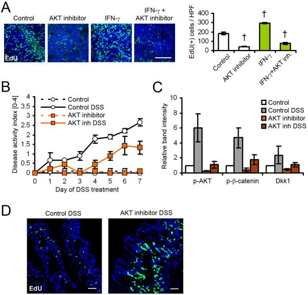
Figure 4.
Epithelial cell proliferation and Dkk1 expression are controlled by AKT. (A) T84 cell proliferation in the presence of AKT inhibitor triciribine and IFN-γ was determined by EdU incorporation for 2 hours following treatment for 22 hours. Inhibition of AKT signaling attenuated IFN-γ-induced proliferation. Scale bar, 100μm. (B) AKT inhibition ameliorated intestinal inflammation in vivo. The graph shows the results from three mice per group. (C) Immunoblot analysis of mucosal lysates showed strongly inhibited AKT and β-catenin activity, as well as Dkk1 expression, in mice receiving DSS and AKT inhibitor. (D) EdU incorporation for 2 hours revealed increased crypt cell proliferation in animals treated with triciribine. Scale bars, 50μm. Data in all graphs in this figure are represented as mean ± s.e.m. † P < 0.01 vs control (ANOVA with Dunnett’s post-test). For additional information please see also Figure S3 in the supplemental material.

