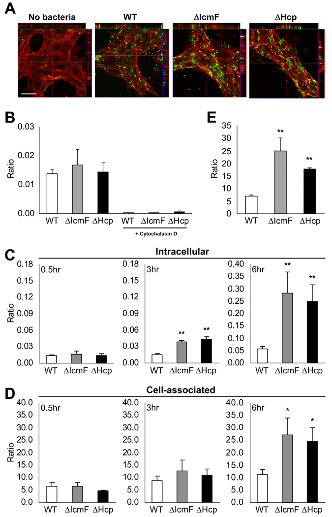
Figure 2. T6SS Mutants Display Higher Intracellular and Cell-associated Accumulation in MODE-K cells.
(A) Confocal image of bacteria inside MODE-K cells. WT, ΔIcmF, or ΔHcp was incubated with MODE-K for 6hr. MODE-K cells were rinsed with PBS, fixed in 4% PFA, and stained for H. hepaticus (green) and the eukaryotic cell membrane marker wheat germ agglutinin (red). Scale bar represents 30µm. See also Movie S1.
(B) Cytochalasin D inhibits uptake of H. hepaticus. Prior to incubation with bacteria, MODE-K cells were treated with 10µM cytochalasin D for 1hr. Bacteria were added at an MOI of 100. After 0.5hr incubation at 37°C under mi croaerophilic conditions, cells were treated with 100µg/ml gentamicin, and intracellular bacteria plated for enumeration. Results are expressed as colony-forming units (CFUs) of intracellular bacteria divided by number of MODE-K cells. Error bars indicate SEM from 3 experiments.
(C, D) Gentamicin protection assay in which MODE-K cells were incubated with bacteria at an MOI of 100. After 0.5, 3, or 6hr incubation, media was replaced with 100µg/ml gentamicin for enumeration of intracellular bacteria (C) or without gentamicin for cell-associated bacteria (D). Cells were washed and bacteria plated for quantification. Ratios are expressed as CFUs of bacteria divided by number of MODE-K cells. Error bars indicate SEM from 3–5 experiments. *p<0.05, **p<0.01 vs WT.
(E) Increased adherence of T6SS mutants is not dependent on bacterial internalization. Prior to co-culture, MODE-K cells were treated with 10µM cytochalasin D. Bacteria were added at an MOI of 100 for 6hr at 37°C under microa erophilic conditions. Bacteria were plated for enumeration. Results are expressed as CFUs of bacteria divided by number of MODE-K cells. Error bars indicate SD from 2 experiments. **p<0.01 vs WT. See also Figure S1.

