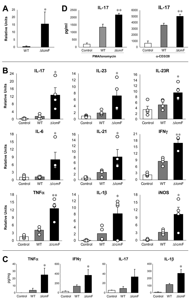
Figure 5. T6SS Mutant Leads to Greater Inflammation in the Colon in the Rag Adoptive Transfer Model of Colitis.
(A) ΔIcmF-colonized animals have greater amounts of H. hepaticus 16S rRNA in the colon compared to WT-colonized animals. RNA was collected from colons. Levels of 16S were quantified by qRT-PCR. Data are representative of 3 experiments, with n=4 per group. Error bars show SEM. *p<0.05 vs WT.
(B) RNA from the colon was analyzed by qRT-PCR for inflammatory cytokines. Experimental values were normalized against L32. Results are representative of 5 experiments, with n=4 per group. Each circle represents an individual animal. Error bars show SEM. *p<0.05, **p<0.01 vs WT. See also Figure S3.
(C) Colons were cultured ex vivo for 24hrs. Supernatants were assayed for cytokine by ELISA. Samples were normalized to total protein. Data are representative of 2 experiments, with n=4 per group. Each circle represents an individual animal. Error bars show SEM. *p<0.05 vs WT.
(D) Mesenteric lymph nodes pooled from each experimental group were restimulated with either PMA and ionomycin, or α-CD3 and α-CD28 for 24hrs. Supernatants were assayed by ELISA. Results are from 2 experiments, with n=4 mice per group. Error bars show SEM. **p<0.01 vs WT.

