Fig. 2.

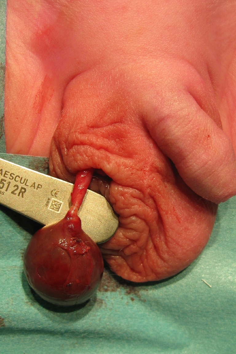
Clear demonstration of the point of torsion in the spermatic cord: Cremasteric tissues, internal spermatic fascia, and tunica vaginalis all seem involved. The testis is still covered by tunica vaginalis

Clear demonstration of the point of torsion in the spermatic cord: Cremasteric tissues, internal spermatic fascia, and tunica vaginalis all seem involved. The testis is still covered by tunica vaginalis