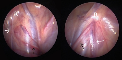
Fig. 4.
Typical laparoscopic image at the age of 5 of the internal inguinal rings in a patient with left-sided PTT. The black arrow indicates the vas crossing the iliac vessels and then ending blindly close to the closed internal inguinal ring (R), the level to which the torsion of the cord was transmitted. The white arrow shows the atrophic testicular vessels or their fibrotic remnants running toward the internal inguinal ring. Normal contralateral situation

