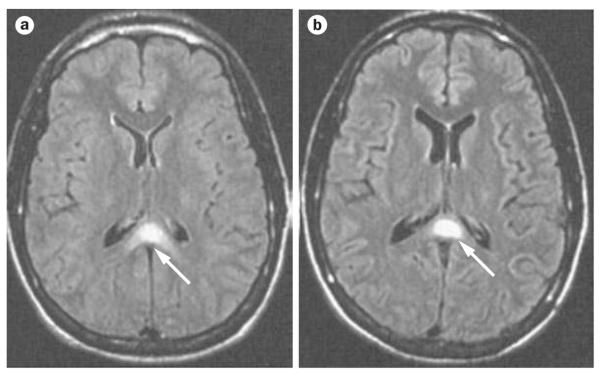
Figure 2.
FLAIR images of the brain of a patient with cerebral malaria. a | Loss of sulci and narrowing of the ventricles (brain swelling), with hyperintensity in the semiovale centrum along with abnormal signal in the splenium of the corpus callosum (arrow). b | One week after the onset of illness, the image shows widening of the sulci and ventricles, with resolution of hyperintensities, except for the lesion in the splenium of the corpus callosum (arrow). Abbreviation: FLAIR, fluid-attentuated inversion recovery. Permission obtained from the American Society of Neuroradiology © Cordoliani Y. S. et al. AJNR Am. J. Neuroradiol. 19, 871–874 (1998).

