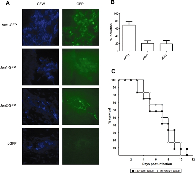
Fig. 7.

Expression of Jen1-GFP and Jen2-GFP in Candida albicans cells infecting the kidney. A.C. albicans cells infecting the kidneys of mice were visualized by staining with Calcofluor white (CFW), and then those that expressed their GFP fusions were imaged by fluorescence microscopy (GFP): Act1-GFP, Jen1-GFP and Jen2-GFP. B.The proportion of C. albicans cells infecting the kidney that display GFP fluorescence above background levels. C.Comparison of the virulence of wild-type and jen1jen2 cells in the mouse model of systemic candidiasis.
