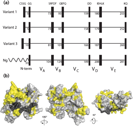
Fig. 2.
(a) Schematic representation of fHbp, showing positions of blocks of invariant residues (shown as black vertical rectangles). The top three panels show representative architectures of three N. meningitidis fHbp variants in groups 1, 2 and 3 (peptide ID nos 1, 16 and 28, respectively). The amino acid positions of the last residue in each variable segment are shown. With the exception of a longer, unrelated, amino-terminal element, two N. gonorrhoeae orthologues (Ng, GenBank accession nos AE004969 and CP001050) had the identical six invariant blocks of residues that flanked segments VA to VE. (b) Space-filling structural models of fHbp based on the coordinates of the protein in a complex with a fragment of human fH (Schneider et al., 2009). The light-grey residues represent the variable amino acids located within the modular variable segments. The invariant blocks of residues separating each of the variable segments are shown in dark grey. The yellow residues represent the invariant amino acids outside of these blocks. The model on the left is the surface predicted to be anchored to the cell wall. The model in the centre has been rotated 18 ° on the y axis from the corresponding model on the left, while the model on the right has been rotated 9 ° on the x axis as compared with the model in the centre. The figure was constructed with PyMol (http://www.pymol.org).

