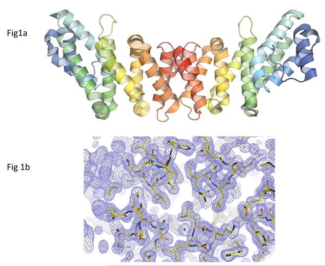
Figure 1.
Crystal structure of D. rerio MIF4Gdb. (A) Ribbon diagram of D. rerio MIF4Gdb structure. A rainbow gradient is used to color each polypeptide chain from its N-terminus (blue) to C-terminus (red). Pairs of α-helices can be seen to form an extended sheet. The C-termini form dimer contacts and the N-terminal regions are available for interaction with other proteins, in keeping with other homologs. (B) Representative lectron density of the MIF4Gdb structure. Amino acid residues are colored by atom types (carbon: green; oxygen: red; nitrogen: blue), and the 2Fobs - Fcalc map is contoured at 1.5σ.

